Crush Injury And
Hyperbaric Oxygen Therapy (HBOT)
Crush injury is directly associated with trauma while skeletal muscle compartment syndromes arise from ischemia, venous outflow obstruction, exertion, external compression as well as trauma. They have the following in common:
- Ischemia and hypoxia at the injury site
- A gradient of injury
- The potential for self-perpetuation of the injury
Management of the most severe presentations of these conditions almost always requires surgery. Hyperbaric oxygen (HBO2) is an effective intervention that counteracts the pathophysiological events which occur with these conditions.
Studies show statistically significant reductions in the loss of muscle function, metabolites associated with muscle injury, edema, and muscle necrosis when HBO2 is used in crush injury, compartment syndrome models.
Consequently, HBOT should be used as a therapeutic adjunct for these conditions when their severity makes expectations of complications and/or less than optimal outcomes likely with usual surgical and medical interventions.121

Crush Injury
By James R. Dickson M. D., FACEP
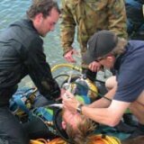
Crush injuries are rarely encountered in everyday in trauma care but become an important injury in disaster situations. These injuries are commonly found in victims of collapsed structures, which may result from earthquakes, hurricanes, tornados, bombings, and other largescale events.
It is critically important that medical and rescue personnel coordinate their efforts in caring for victims of crush injuries. Serious morbidity and even mortality may occur if a crushed victim does not receive medical care until after extrication by rescue personnel. Aggressive medical treatment of the patient before and during extrication will help prevent renal and cardiac complications of the injury.239 Without early treatment, patients may die immediately upon extrication or days to weeks later from complications. The cause of these deaths and complications is crush injury syndrome, the systemic manifestations of muscle crush injury and cell death.
History
Recognition of the medical consequences of crush injuries dates back to World War II.240 Reports of the treatment of crush injury were published after the disasters described below:
July 28, 1976 · Tangshan, China
Earthquake - magnitude 7.8 on the Richter scale. 242,769 died; 164,851 injured. Crush injury syndrome occurred in 2% to 5% of injured people. One woman trapped in a collapsed building survived for 13 days.
- Findings:
- Patients with a history of being crushed for a few hours must be observed carefully for crush injury syndrome, as they often appeared initially to have only minimal injury.
- The area crushed and duration of being buried did not predict the occurrence of acute renal failure.241
November 11, 1982 · Tyre, Lebanon
Eight-story building collapse. 100 people buried; 20 survivors extricated in the subsequent 28 hours, 8 with crush injury syndrome.
- Findings:
- Seven of the eight patients with severe crush injury syndrome received aggressive treatment. None required dialysis, and all but one survived.
- The patient who did not receive aggressive treatment developed acute renal failure requiring hemodialysis.239
December 7, 1988 · Armenia, Former Soviet Union
Earthquake magnitude 6.9 on the Richter scale. More than 100,000 injured; 15,254 extricated alive from the rubble. Little to no care or resuscitation was given to victims prior to arrival at a hospital.
- Findings:
- Crush injury was the third most frequent injury, but was the leading cause of death.
- In many cases, further deterioration was noted upon release of crushed extremities.
- Acute renal failure and the need for dialysis were common.242
January 17, 1995 Kobe, Japan
Earthquake magnitude 7.2 on the Richter scale 41,000 injured; 5,000 died. Few, if any, patients had early treatment.
- Findings:
- 54% of victims developed acute renal failure. These patients had a longer interval between rescue and initiation of treatment.
- Only 11% of patients who received more than 6 L of fluid a day developed renal failure.243, 247
Pathophysiology
Mechanisms of Muscle Cell Injury
The pathophysiology of crush injury begins with muscle injury and muscle cell death.244 Initially three mechanisms are responsible for the death of muscle cells:
- Immediate cell disruption: The local force of the crush causes immediate cell disruption (lysis). Although the effects are immediate, it is probably the least important mechanism.
- Direct pressure on muscle cells: The direct pressure of the crush injury causes the muscle cells to become ischemic. The cells then switch to anaerobic metabolism, generating large amounts of lactic acid. Prolonged ischemia then causes the cell membranes to leak.245 This process occurs during the first hour after the injury.
- Vascular compromise: The force of the injury compresses large vessels, resulting in loss of blood supply to muscle tissue. Normally, muscle can withstand approximately 4 hours without blood flow (warm ischemia time) before cell death occurs. After this time, cells begin to die as a result of vascular compromise.
Release of Substances from Injured Muscle
The mechanisms listed above cause the injured muscle tissue to generate and release a number of substances that may be toxic in the general circulation. The crushing force actually serves as a protective mechanism, preventing these toxins from reaching the central circulation. Once the patient is extricated and the force is released, the toxins are free to travel in the circulation to exert their effects systemically. They can affect organs far from and not involved in the local crush injury. The toxin leak may continue for as long as 60 hours after release of the injury.239 Some of these substances and their consequences are listed below:
- Amino acids and other organic acids contribute to acidosis, aciduria, and dysrhythmia.
- Creatine phosphokinase (CPK) and other intracellular enzymes serve as laboratory markers for crush injury.
- Free radicals, superoxides, peroxides formed when oxygen is reintroduced into ischemic tissue, caus ing further tissue damage.
- Histamine vasodilation, bronchoconstriction.
- Lactic acid major contributor to acidosis and dysrhythmias.
- Leukotrienes lung injury (adult respiratory distress syndrome [ARDS]), and hepatic injury.
- Lysozymes - cell-digesting enzymes that cause further cellular injury.
- Myoglobin precipitates in kidney tubules, especially in the setting of acidosis with low urine pH; leads to renal failure.246
- Nitric oxide causes vasodilation, which worsens hemodynamic shock.
- Phosphate hyperphosphatemia causes precipitation of serum calcium, leading to hypocalcemia and dysrhythmias.
- Potassium hyperkalemia causes dysrhythmias, especially when associated with acidosis and hypocalcemia.
- Prostaglandins vasodilation, lung injury.
- Purines (uric acid) may cause further renal damage (nephrotoxic).
- Thromboplastin disseminated intravascular coagulation (DIC).
There is no correlation between toxic levels of substances such as potassium or myoglobin and the severity of the crush injury or the length of time that the patient was entrapped.247
Other Consequences of Reperfusion
Third Spacing. Leaking cell membranes and capillaries cause intravascular fluids to accumulate in injured tissue. This leads to significant hypovolemia and eventually hypovolemic shock.245, 248 Loss in calcium into injured tissue also contributes to hypocalcemia.
Compartment Syndrome. Muscle groups are surrounded by tough layers of fascial tissue that form compartments. When the muscle tissue in these compartments swells, the pressure within the compartment increases. This leads to worsening of ischemia and further muscle damage. Also, any blood vessels or nerves that travel through that compartment will be injured.
Assessment
The patient with crush injury may present initially with few signs or symptoms.249 Medical personnel must maintain a high index of suspicion in treating victims of crush injury. It may be too late for optimal outcome if treatment is delayed while waiting for signs and symptoms to appear after the limb(s) are released. Crush injury syndrome should be anticipated, so treatment should begin before extrication.
Crush injury syndrome should be suspected in patients with certain patterns of injury. Most patients in whom the syndrome develops have an extensive area of involvement such as a lower extremity and/or the pelvis. It requires more involvement than just one hand or foot. Also, the crushing force must be present for some time before crush injury syndrome can occur. The syndrome may develop after 1 hour in a severe crush situation, but usually it takes 4 to 6 hours of compression for the processes that cause crush injury syndrome to take place.
Signs and Symptoms of Crush Injury
Some or all of the following may be present:
- Skin injury may be subtle
- Swelling usually a delayed finding
- Paralysis may cause crush injury to be mistaken as a spinal cord injury
- Paresthesias, numbness may mask the degree of damage
- Pain often becomes severe upon release
- Pulses distal pulses may or may not be present
- Myoglobinuria the urine may become dark red or brown, indicating the presence of myoglobin
Hyperkalemia
As mentioned earlier, hyperkalemia is often present in a patient with crush injury syndrome. In the absence of laboratory analysis, the degree of hyperkalemia can be estimated crudely from the electrocardiogram (ECG). It is most helpful to compare serial ECGs than to look at only one. The classic electrocardiographic changes are as follows:
- Mild hyperkalemia (5.56.5 mEq/L) peaked T waves
- Moderate hyperkalemia (6.57.5 mEq/L) prolonged PR interval, decreased P wave amplitude, depression or elevation of ST segment, slight widening of the QRS complex.
- Severe hyperkalemia (7.58.5 mEq/L) further widening of the QRS due to bundle branch or intraventricular blocks, flat and wide P waves, Wenckebach, ventricular ectopics.
- Life-threatening hyperkalemia (>8.5 mEq/L) loss of P waves; AV blocks; ventricular dysrhythmias; further widening of the QRS complex, eventually forming a sinusoid pattern.
Compartment Syndrome
As mentioned under pathophysiology, compartment syndrome may coexist with crush injury. Signs and symptoms associated with this include the following:
- Severe pain in the involved extremity
- Pain on passive stretching of the muscles involved
- Decreased sensation in branches of the involved peripheral nerves
- Elevated intracompartmental pressures on direct manometry
Treatment
General
The victim of crush injury should be treated initially as any other multiple trauma victim, according to the various trauma life support course guidelines. The airway must be secured and protected from dust impaction. Adequate ventilation must be ensured and maintained along with adequate oxygenation. In a disaster situation with limited supplies, it may be prudent to conserve oxygen by using the lowest flow rate necessary to adequately oxygenate the patient, as indicated by oxygen saturation measurements and clinical assessment. Circulation must be supported and shock aggressively treated.
As mentioned earlier, it is critically important to coordinate with rescue personnel so that medical treatment can begin prior to the patients release.
Intravenous Fluid
The mainstay of treatment for crush injury syndrome is intravenous fluid.245 Initially, any preexisting dehydration or fluid loss should be corrected. The patients fluid volume status should be corrected to normal, which may require several liters of fluid, before any compression is lifted from the patient. Multiple intravenous lines are appropriate, because the patient will require large fluid volumes and there is a risk of dislodgement during extrication. Intravenous (IV) fluids containing potassium (e.g., lactated Ringer's solution) should be avoided. Normal saline is a good initial choice.
Once the compression is lifted, it is critical to maintain a high urine output. Placement of a Foley catheter will allow more accurate measurements of urine output as well as pH. One formula that can be used to maintain an alkaline urine output of 8 L/d is the infusion of 12 L/d of Normal Saline Solution (NSS) with 50 mEq of sodium bicarbonate per liter of fluid, plus 120 grams of mannitol daily to maintain this urine output. After the building collapse in Tyre, Lebanon, an average of 568 ml/h of IV fluid was given to the victims to successfully prevent renal failure.239 Another regimen is 12 L/d (500 ml/h) of a solution containing sodium, 110 mmol/L; chloride, 70 mmol/L; bicarbonate, 40 mmol/L; and mannitol, 10 gm/L.250
Sodium Bicarbonate
Sodium bicarbonate has seve ral actions that are useful in the patient with crush injury syndrome. It will reverse the preexisting acidosis that is often present. It is one of the first steps in treating hyperkalemia. It will also increase the urine pH, thus decreasing the amount of myoglobin precipitated in the kidneys.
It is recommended that 50 to 100 mEq of bicarbonate, depending on severity of injury, be given to a victim prior to release from compression. This may be followed by continuous infusions of bicarbonate, as described under intravenous fluids.
Treatment of Hyperkalemia
In addition to sodium bicarbonate, other treatments may be needed to reverse hyperkalemia, depending on the severity of injury:
- Insulin and glucose
- Calcium intravenously for life-threatening dysrhythmias
- Beta-2 agonists albuterol, metaproterenol sulfate (Alupent), etc...
- Potassium-binding resins such as sodium polystyrene sulfonate (Kayexalate)
- Dialysis, especially in patients with acute rena l failure.251,252
Alkaline Diuresis
The patient with crush injury syndrome should maintain a urine output of at least 300 ml/h with a pH higher than 6.5.239 This can be achieved with intravenous fluids, mannitol, and sodium bicarbonate (44 to 50 mEq/liter of intravenous fluid, as described above ). Other treatments that have been used are renal-dose" dopamine at 2 to 5 mg/kg/min and furosemide at 1 mg/kg. Acetazolamide, 250 to 500 mg, may be used if the patient becomes too alkalotic.
Intravenous Mannitol
Intravenous mannitol has several beneficial actions in the victim of crush injury.248 It protects the kidneys from the effects of rhabdomyolysis, increases extracellular fluid volume, and increases cardiac contractility. In addition, intravenous administration of mannitol for 40 minutes successfully treated compartment syndrome, with relief of symptoms and reduc tion of swelling.245, 248
Mannitol can be given in doses of 1 gm/kg or added to the patient's intravenous fluid as a continuous infusion. The maximum dose is 200 gm/d; doses higher than this can cause renal failure. Mannitol should be given only after good urine flow has been established with IV fluid.
Wound Care
Wounds should be cleaned, debrided, and covered with sterile dressings in the usual fashion. Splinting the limb at heart level will help to limit edema and maintain perfusion. Intravenous antibiotics are used frequently. Medications for pain control can be given as appropriate. Tourniquets are controversial and usually not necessary.
Pneumatic Antishock Garment (PASG)
Application of the PASG should be avoided. The use of PASG has been reported to cause compartment syndrome and crush injury syndrome.253
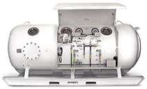
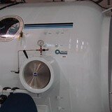
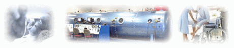
Hyperbaric Oxygen
There are case reports of hyperbaric oxygen improving the outcome of victims of crush injury.254, 255 The use of this modality will be limited in disaster situations because of lack of access to hyperbaric chambers.
Amputation
Amputation in the field should be used only as a last resort. It might be the appropriate rescue strategy for a patient whose life is in immediate danger from another collapse or hazardous materials and who cannot be extricated by any other means. It is a difficult field procedure that greatly increases the patient's risk of infection and bleeding.
Fasciotomy
Field fasciotomy is also a controversial procedure, which may further expose the patient to the risk of infection and bleeding.249 It converts a closed injury to an open one, risking infection and sepsis. Several studies indicate a worse outcome in patients who received fasciotomy compared with those who did not.245, 249 It is felt to be indicated only in a potentially viable extremity that is pulseless due to elevated intracompartmental pressure.256 Fasciotomy was felt to be useful in preventing cases of Volkmans ischemic contracture after the Tangshan earthquake.241 In Israel, fasciotomy was reserved as a treatment of last resort in cases refractory to the use of intravenous mannitol.245
When used as an adjunct to orthopedic surgery and antibiotics, hyperbaric oxygen therapy (HBOT) shows promise as a way to decrease complications from severe crush injuries. HBOT increases oxygen delivery to the injured tissues, reduces swelling and provides an improved environment for healing and fighting infection.